Image BankNews media are welcome to download and reproduce the following images in the context of explaining neuromodulation therapies to the public. Photo credits are required where indicated, and some links to sources provided.
Media Contact Neuromodulation Images
Credit (required): Photo courtesy of Duke University Click on image to download a 300 dpi jpg image For larger images, contact Duke Photography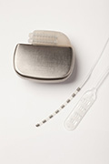
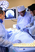
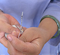
Caption: In spinal cord stimulation, a rounded wire is used for a temporary trial to test a patient’s response to the therapy, and either a rounded wire or paddle-shaped lead is used for a permanent implantation. (2013) Credit (required): Photo courtesy of Duke University Click on image to download a 300 dpi jpg image For larger images, contact Duke Photography
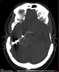
Caption: This CT scan image depicts closed-loop responsive neurostimulation, which is undergoing clinical trials for halting seizure activity in epilepsy. Click on image to download a 300 dpi jpg image
Credit: Image courtesy of the North American Chapter of the International Neuromodulation Society (NANS/INS) (2012) Click on image to download a 300 dpi jpg image
Credit: Image courtesy Annu Navani, M.D. Click on image to download a jpg file
Credit: Image courtesy Magdy Hassouna, M.D., Ph.D. Click on image to download a 300 dpi jpg image 
Caption: International Neuromodulation Society member Dr. Peter Staats, an interventional pain specialist, inserts the lead of a spinal cord stimulation system along the spine of a chronic pain patient. Credit: Image courtesy of St. Jude Medical (2012) Click on image to download a larger jpg image 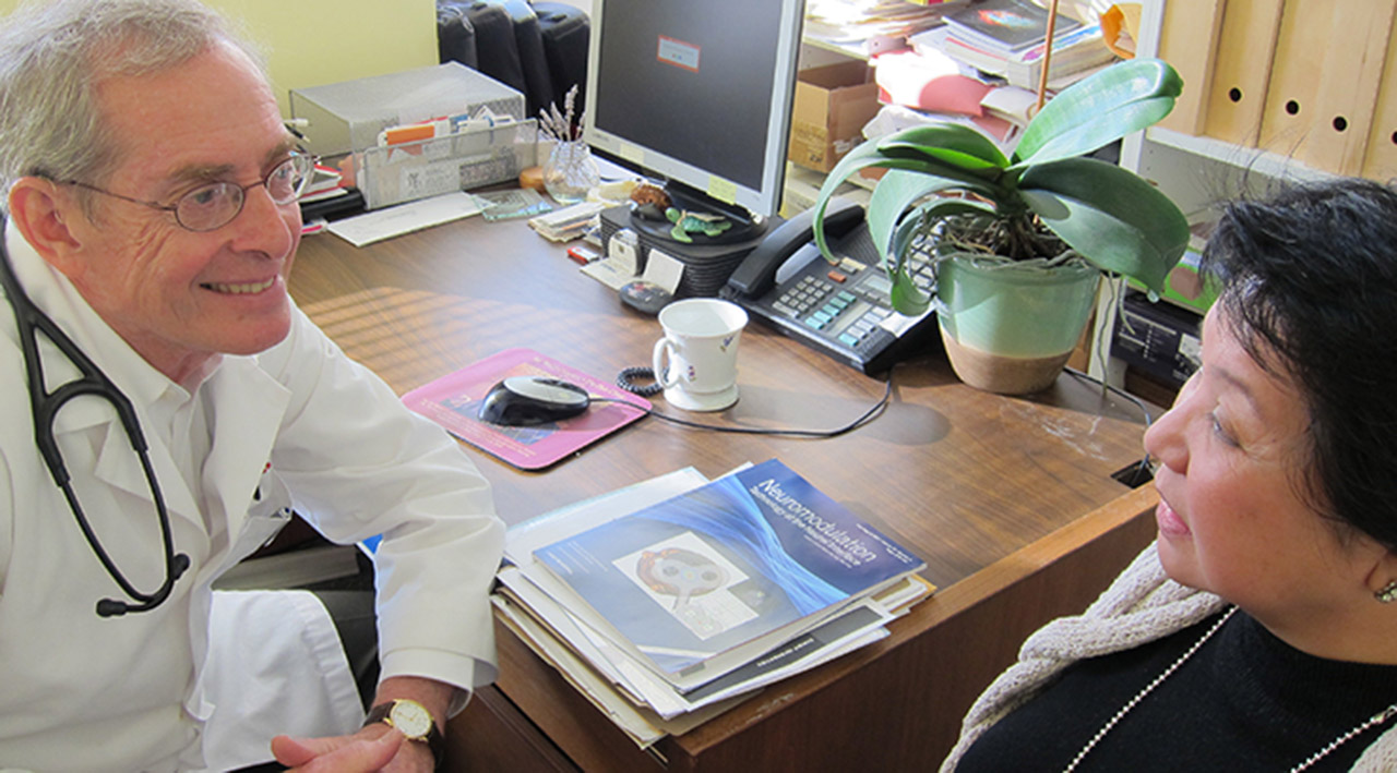
Caption: Dr. Elliot Krames speaks with a patient at the Pacific Pain Treatment Center he founded in San Francisco. Credit: Image courtesy of the International Neuromodulation Society (INS) (2013) Click on image to download a larger jpg image 
Caption: Cochlear implants were an early application of neuromodulation. Credit: Image courtesy of the International Neuromodulation Society (INS) (2012)
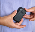
Caption: A demonstration patient controller is shown in a clinical examination room. The controller is held near the implantable pulse generator to turn it on or off or switch between stimulation programs that have been preset for the patient. (The pacemaker-like pulse generator is often implanted on the upper buttock, or the chest wall or abdomen, depending on the neurostimulation treatment, such as spinal cord stimulation, sacral neuromodulation for some genitourinary conditions, or deep brain stimulation to manage tremor from movement disorder.) Credit: Image courtesy of the International Neuromodulation Society (INS) (2012) Click on image to download a 300 dpi jpg image
Credit: Image courtesy of the International Neuromodulation Society (INS) (2012) Click on image to download a 300 dpi jpg image
Credit (required): Image courtesy of Dr. Kaare Meier, M.D. Ph.D., Department of Neurosurgery, Aarhus University Hospital, Denmark (2015) (Permission for use limited to any non-commercial purpose, with proper credit and no alteration.) Click on image to download a 300 dpi jpg image
Credit (required): Image courtesy of Dr. Kaare Meier, M.D. Ph.D., Department of Neurosurgery, Aarhus University Hospital, Denmark (2013) (Permission for use limited to any non-commercial purpose, with proper credit and no alteration.) Click on image to download a 300 dpi jpg image
(Permission for use limited to any non-commercial purpose, with proper credit and no alteration.) Click on image to download a 300 dpi jpg image
Credit (required): Image courtesy of Dr. Kaare Meier, M.D. Ph.D., Department of Neurosurgery, Aarhus University Hospital, Denmark (2015) (Permission for use limited to any non-commercial purpose, with proper credit and no alteration.) Click on image to download a 300 dpi jpg image Credit (required): Image courtesy of Dr. Kaare Meier, M.D. Ph.D., Department of Neurosurgery, Aarhus University Hospital, Denmark (2015) (Permission for use limited to any non-commercial purpose, with proper credit and no alteration.) Click on image to download a 300 dpi jpg image
Credit (required): Image courtesy of Dr. Kaare Meier, M.D. Ph.D., Department of Neurosurgery, Aarhus University Hospital, Denmark (2015) (Permission for use limited to any non-commercial purpose, with proper credit and no alteration.) Click on image to download a 300 dpi jpg image
Credit (required): Image courtesy of Dr. Kaare Meier, M.D. Ph.D., Department of Neurosurgery, Aarhus University Hospital, Denmark (2015) (The image may be reproduced for any non-commercial purpose, as long as there is proper credit given and the picture is not altered.) Click on image to download a 300 dpi jpg image
Credit (required): Image courtesy of Dr. Kaare Meier, M.D. Ph.D., Department of Neurosurgery, Aarhus University Hospital, Denmark (2015) (The image may be reproduced for any non-commercial purpose, as long as there is proper credit given and the picture is not altered.) Click on image to download a 300 dpi jpg image
Credit (required): Image courtesy of Dr. Kaare Meier, M.D. Ph.D., Department of Neurosurgery, Aarhus University Hospital, Denmark (2015) (The image may be reproduced for any non-commercial purpose, as long as there is proper credit given and the picture is not altered.) Click on image to download a 300 dpi jpg image
Credit (required): Image courtesy of Dr. Kaare Meier, M.D. Ph.D., Department of Neurosurgery, Aarhus University Hospital, Denmark (2015) (The image may be reproduced for any non-commercial purpose, as long as there is proper credit given and the picture is not altered.) Click on image to download a 300 dpi jpg image
Credit (required): Image courtesy of Dr. Kaare Meier, M.D. Ph.D., Department of Neurosurgery, Aarhus University Hospital, Denmark (2015) (The image may be reproduced for any non-commercial purpose, as long as there is proper credit given and the picture is not altered.) Click on image to download a 300 dpi jpg image
Credit (required): Image courtesy of Dr. Kaare Meier, M.D. Ph.D., Department of Neurosurgery, Aarhus University Hospital, Denmark (2015) (The image may be reproduced for any non-commercial purpose, as long as there is proper credit given and the picture is not altered.) Click on image to download a 300 dpi jpg image
Credit (required): Image courtesy of Dr. Kaare Meier, M.D. Ph.D., Department of Neurosurgery, Aarhus University Hospital, Denmark (2015) (The image may be reproduced for any non-commercial purpose, as long as there is proper credit given and the picture is not altered.) Click on image to download a 300 dpi jpg image
Credit (required): Image courtesy of Dr. Kaare Meier, M.D. Ph.D., Department of Neurosurgery, Aarhus University Hospital, Denmark (2015) (The image may be reproduced for any non-commercial purpose, as long as there is proper credit given and the picture is not altered.) Click on image to download a 300 dpi jpg image 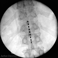
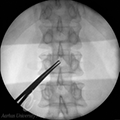
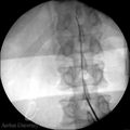
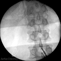
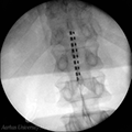
Caption: This fluoroscopy image shows the lead placed in the midline inside the epidural space. The tip (contact #1) is at the level of the intervertebral disc Th11/Th12. Credit (required): Image courtesy of Dr. Kaare Meier, M.D. Ph.D., Department of Neurosurgery, Aarhus University Hospital, Denmark (2015) (The image may be reproduced for any non-commercial purpose, as long as there is proper credit given and the picture is not altered.) Click on image to download a 300 dpi jpg image
Credit (required): Image courtesy of Dr. Kaare Meier, M.D. Ph.D., Department of Neurosurgery, Aarhus University Hospital, Denmark (2015) (The image may be reproduced for any non-commercial purpose, as long as there is proper credit given and the picture is not altered.) Click on image to download a 300 dpi jpg image
Credit (required): Image courtesy of Dr. Kaare Meier, M.D. Ph.D., Department of Neurosurgery, Aarhus University Hospital, Denmark (2015) (The image may be reproduced for any non-commercial purpose, as long as there is proper credit given and the picture is not altered.) Click on image to download a 300 dpi jpg image
Credit (required): Image courtesy of Dr. Kaare Meier, M.D. Ph.D., Department of Neurosurgery, Aarhus University Hospital, Denmark (2015) (The image may be reproduced for any non-commercial purpose, as long as there is proper credit given and the picture is not altered.) Click on image to download a 300 dpi jpg image Brain Images
Credit: Image courtesy Mark Dow, University of Oregon. Although the image is in the pubic domain, the creator appreciates a link sent to dow(at)uoregon.edu Click on image to download a larger image. Credit: Image courtesy National Institute of Mental Health. Click on image to download a larger image. Credit: Image courtesy National Institute of Mental Health. Click on image to download a larger (rectangular) image.
Credit: Image courtesy National Institutes of Health Click on image to download a larger image. Credit: Image courtesy National Institute of Mental Health Click on image to download a larger image. Credit: National Library of Medicine

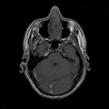
Caption: A sagittal magnetic resonance image (MRI) of the brain. National Science Foundation (NSF)-supported fundamental research led to the development of MRI technology. Credit: Courtesy FONAR Corporation (via NSF). Click on image to download a high-resolution TIFF image. Academic R&D Images
Credit: National Science Foundation Click on image to download a larger image.
Credt: EurekAlert Click on image to download a larger image.
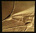
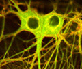
Caption: Electron micrograph of human neurons. Credit: National Institute of Mental Health Click on image to download
Credit: Image courtesy of the National Institutes of Health Click on image to download a larger image
Credit: Image by Tina Carvalho, University of Hawaii at Manoa, courtesy of the National Institute of General Medical Sciences/NIH Click on image to download a larger image Conditions That Are Current, Developing, or Investigative Indications
Credit: Brian T. Gold, Department of Anatomy and Neurobiology, University of Kentucky (via NSF). Click on image to download a a high-resolution TIFF image. Bioelectronic Medicine Images or Tables
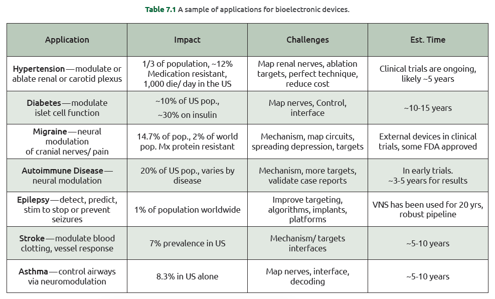
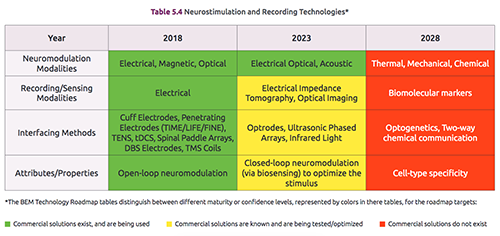
Credit: 2018 Bioelectronic Medicine Technology Roadmap, SRC, Durham, NC, 2018. [Online] Available: https://www.src.org/library/publication/p095388/p095388.pdf Click on image to download a larger png image.
Credit: 2018 Bioelectronic Medicine Technology Roadmap, SRC, Durham, NC, 2018. [Online] Available: https://www.src.org/library/publication/p095388/p095388.pdf Click on image to download a larger png image. Caption: Recording and neurostimulation technologies for bioelectronic medicine from the report "2018 Bioelectronic Medicine Technology Roadmap" by the Semiconductor Research Corporation and NIST. Credit: 2018 Bioelectronic Medicine Technology Roadmap, SRC, Durham, NC, 2018. [Online] Available: https://www.src.org/library/publication/p095388/p095388.pdf Click on image to download a larger png image. |
| Last Updated on Monday, July 17, 2023 07:31 AM |











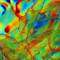
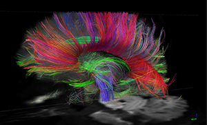
 Caption: Diffusion Spectrum MRI (DSI) of the human brain obtained with the MGH-UCLA Human “Connectom” Scanner acquired in 8 min. Where images of this quality were previously obtained in scans of 1 hr or longer, improved scanner performance coupled with innovations in RF coil design, MRI scanning physics, and mathematics of diffusion MRI reconstruction have reduced the total scan time by 6-fold or more (here from 48 min to 8 min). Most of these innovations will be readily transferable to most or all general-purpose MRI systems, and make possible practical high-resolution diffusion MRI on a routine basis (courtesy Laurence Wald, Van Wedeen). The fiber tracks are color-coded by direction: red=left-right, green=anterior-posterior, blue=through brain stem.